Our adhesion model measures the strength of cell attachment using the controlled detachment assay, and static migration assay. This platform delivers reproductible analysis of cell adhesion dynamics starting with defined ligands through to migration dynamics in extravascular tissue models.

Immune cell profiling
› Cell locomotion (speed and direction)

Drug screening and therapeutic development
› Multi-throughput screening
› Dose-response analysis
› IC50 determination

Adhesion mechanism assessment
› Controlled detachment assay:
• Ligand-specific adhesion analysis
• Recombinant proteins(e.g. ICAM-1, VCAM-1)
• Extracellular matrices (e.g. fibronectin, collagen)
› Static tissue migration assay:
• Disease and tumor coculture models
• Tissue resident leukocyte trafficking analysis

Bio-imaging
› High-resolution imaging of cell adhesion events under flow
› Real-time visualisation of cell-surface interactions
› Adjustable time-course measurements

Cell tracking
› MesenCount and MesenTrack AI-based softwares
› Label-free or immunofluorescence imaging

Flow Cytometry
› Possible – dependent on adhesive interaction
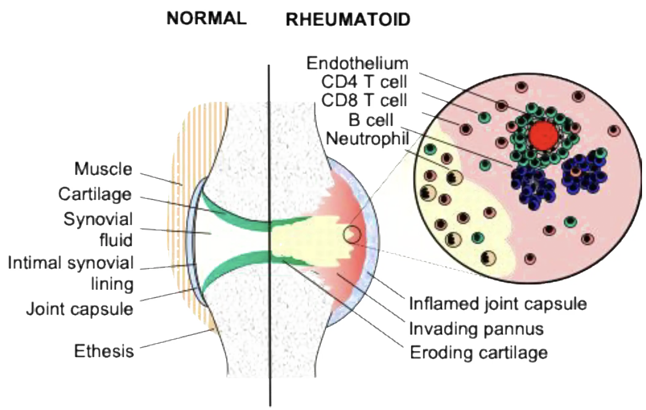
Leukocyte trafficking in the synovium:
migration and retention
Controlled detachment and tissue migration assays represent a comprehensive suite of protocols used to establish how T-cells are retained within complex environments, such as rheumatoid synovial fibroblast cultures.
By applying specific shear stress and using static protocols, we can simultaneously measure adhesion strength, migration speed, and phenotypic impact that are critical factors in understanding leukocyte survival within tumor and chronic inflammatory microenvironments.
These multi-step, in-vitro assessments can provide vital insights into non-vascular processes like pseudoemperipolesis, mediating the integrin activation, stable adhesion and chemotaxis necessary for disease progression.
Example 1
Controlled detachment assays:
Leukocyte binding to surface-bound proteins.
Defining T-cell adhesion in the synovium through shear stress profiling.
Buckley et al. J Immunol 2000; 165:3423-3429
Different adhesion substrate
1) ICAM-1
2) Fibronectin
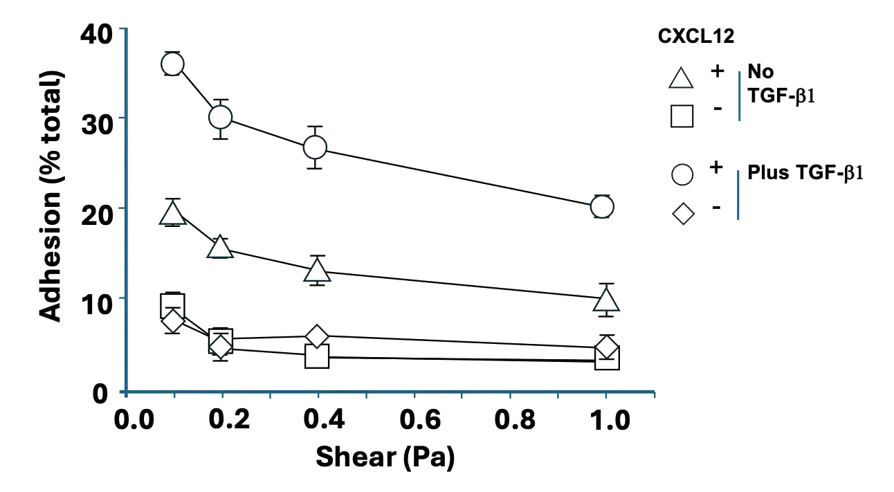
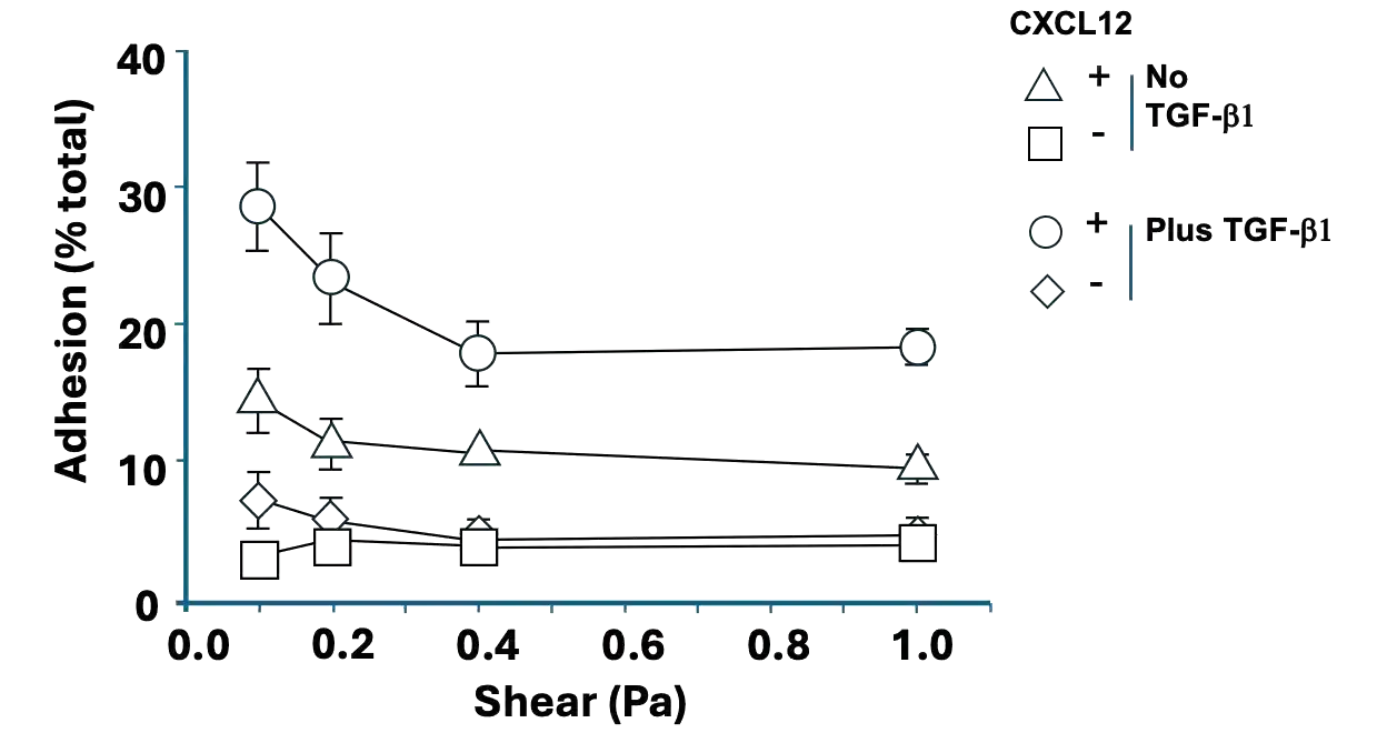
Step-A: TGF-beta isoforms induce functional expression of CXCR4 on short-term CD4 T-cell lines.
The adhesion of CD4 T-cells cultured in the presence of TGF-beta 1 were tested in the presence and absence of CXCL12 on:
1) ICAM-1-Fc protein
2) Fibronectin

Fibronectin
selected
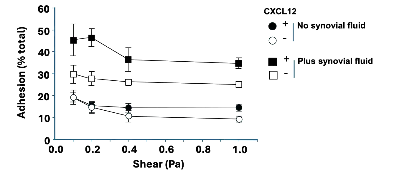
Mesenchymal tissue conditioned
3) Synovial Fluid
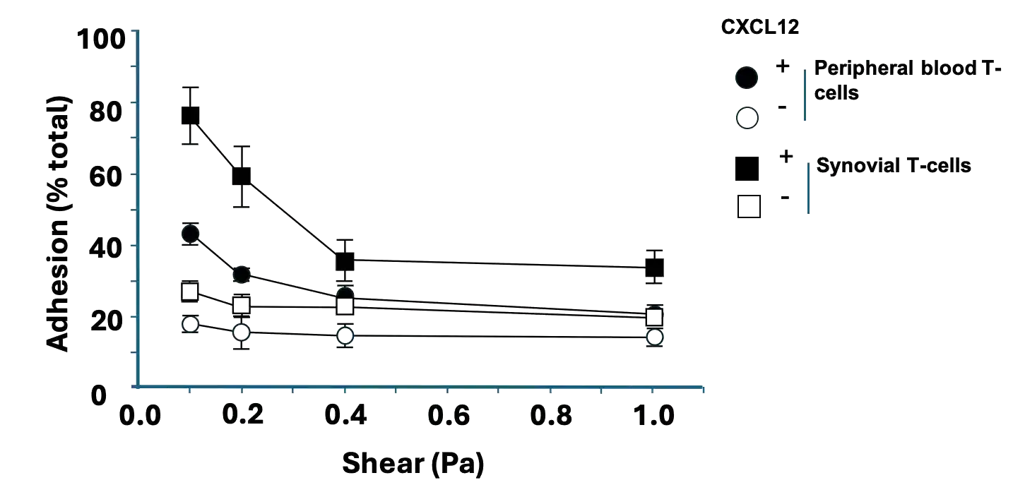
4) Tissue resident T-cells
Step-B: The synovial microenvironment is responsible for the functional up-regulation of CXCR4 on T cells.
3) Synovial T cells cultured with synovial fluid adhered at higher levels compared to synovial T-cells in control medium - further increased by CXCL12.
4) Freshly isolated rheumatoid synovial T-cells adhere to fibronectin at higher levels compared to peripheral blood T-cells from the same patient and were further enhanced by CXCL12.
Example 2
Static tissue migration assays:
Leukocyte trafficking in the extravasculature.
Understanding human T-cell migration in synovial fibroblast cocultures.
Bradfield et al. Arth Rheum; 2003 48:2472-82
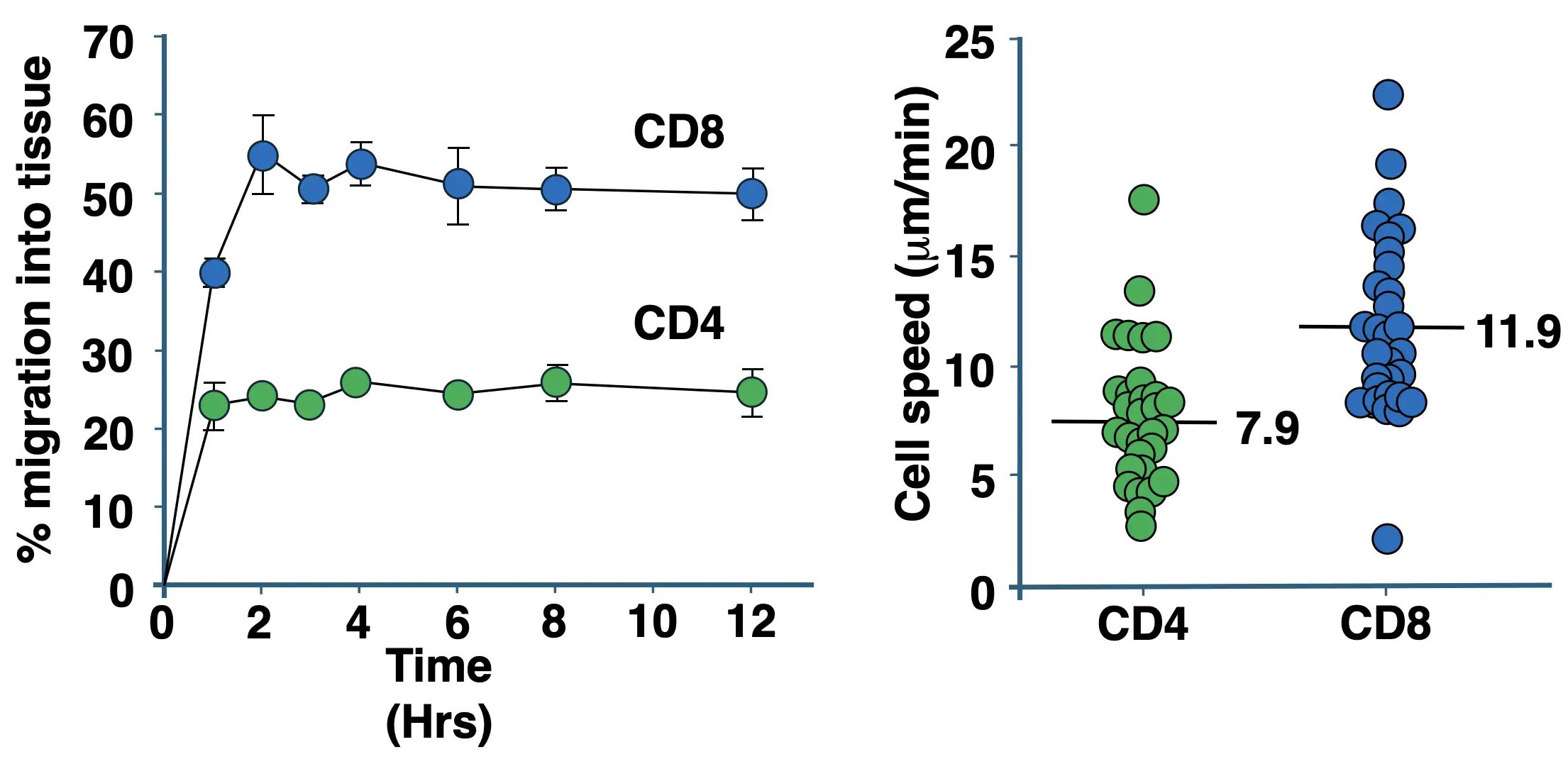
Pseudoemperipolesis of CD4 and CD8 T-cells in rheumatoid fibroblast cocultures. CD4 and CD8 T-cells were co-cultured under static condition with rheumatoid fibroblasts for 12-hrs and the % migration of total cells per unit field expressed as a mean +SD. The speed of migration for CD4 and CD8 T-cells was measured for 2-mins following 2-hrs of culture. For each group, 10 individual cells were tracked in 3 independent experiments and expressed as the median.
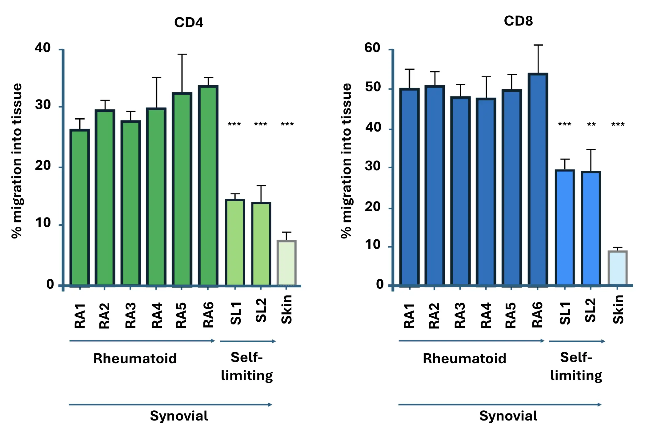
Pseudoemperipolesis of CD4 and CD8 T-cells. CD4 or CD8 T-cells were cocultured with different fibroblasts for 2-hrs. These included six rheumatoid fibroblast cell lines (RA 1–6) as well as synovial fibroblasts derived from patients with self-limiting arthritis (SL1-2), and fibroblasts derived from skin. Significant differences were observed between the pooled rheumatoid panel versus self-limiting arthritis and adult skin for both CD4 and CD8 T-cells.
MesenFlow Technologies SàrlChemin des Aulx 14
1228 Plan-les-Ouates
Geneva, Switzerland
+41 22 32 16 961 (office)
+41 79 36 66 291 (mobile)