

Our flow-based platforms mimic vascular conditions, allowing the study of cellular interactions in dynamic, human-relevant environments.
By combining controlled flow, human cells, real-time imaging, and AI analysis, they enable precise investigation of immune behavior, vascular interactions, and tissue interfaces.
The platform includes two complementary flow systems to cover different levels of biological complexity.
Single-chamber flow system
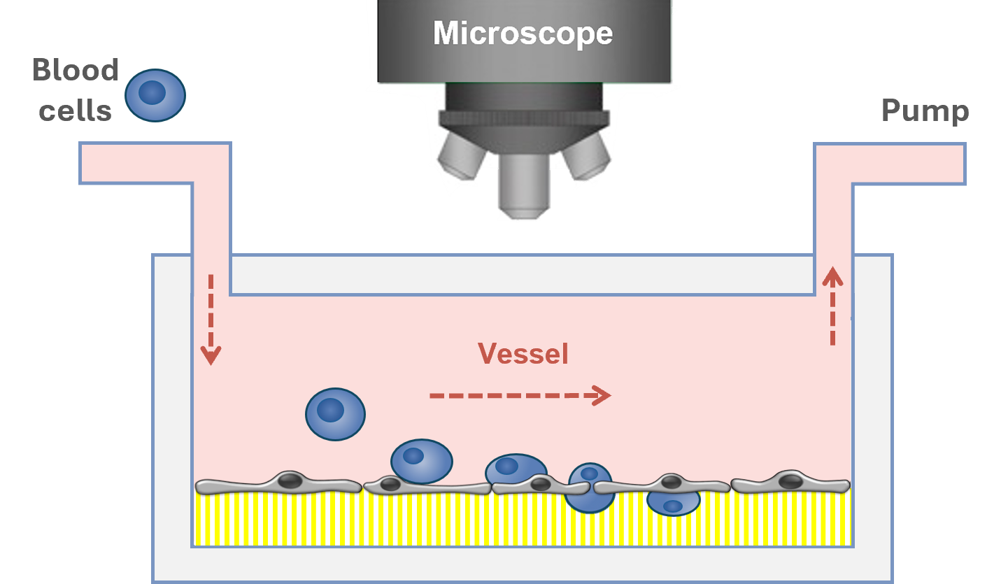
A single-chamber micro-physiological flow platform.
The blood cells, derived from human blood, either as isolated leukocytes or as whole blood, are perfused through a vessel-like channel using a pump system. The flow rate is adjusted to replicate physiological conditions found in blood vessels.
Dual-chamber flow system
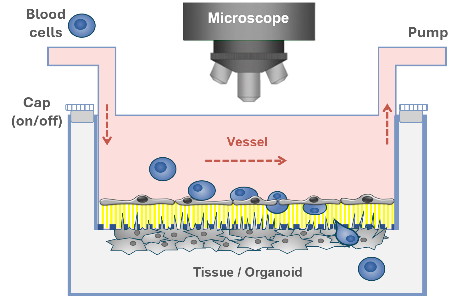
A next-generation dual-chamber platform to study organoids and complex physiological interactions under flow.
Here, a semi-permeable membrane separates the neural tissue compartment from the vascular layer modelling the blood-brain barrier. The platform is designed to be adaptable to a range of 2D tissues and organoid types, enabling the investigation of cell-cell interactions under controlled flow conditions in the vascular chamber which has direct physical contact with the static organoid compartment.
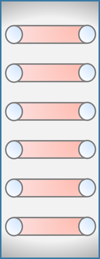
Straight top channel
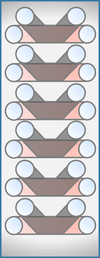
Angled bottom channel
We leverage artificial intelligence (AI) to enhance the analysis of complex cellular behaviors, enabling more accurate and efficient data interpretation. Our AI is applied through proprietary software solutions, developed and continuously refined by our scientists, ensuring accurate and meaningful interpretation of cellular dynamics. Together, these tools represent a shift from manual methods to advanced, label-free AI-driven leukocyte tracking, delivering greater efficiency, consistency and insight.
Precise quantification of leukocytes
Detailed analysis of cell migration, transmigration and locomotion
AI Softwares
Developed by MesenFlow, these software packages were established through deep convolutional neural network algorithm to detect and classify cell transmigration. The algorithm is based on operation blocks that allow the AI to learn patterns it observes within similar cells. High accuracy counts are achieved by extensive data pre-processing, formatting, and post-processing to create a pipeline that integrates well within our current workflow.
Key Advantages

100x faster performance than manual counting

Trained on proprietary datasets

Fluorescence-free image analysis using phase contrast technology

Manual Counting
AI-guided Counting
➔
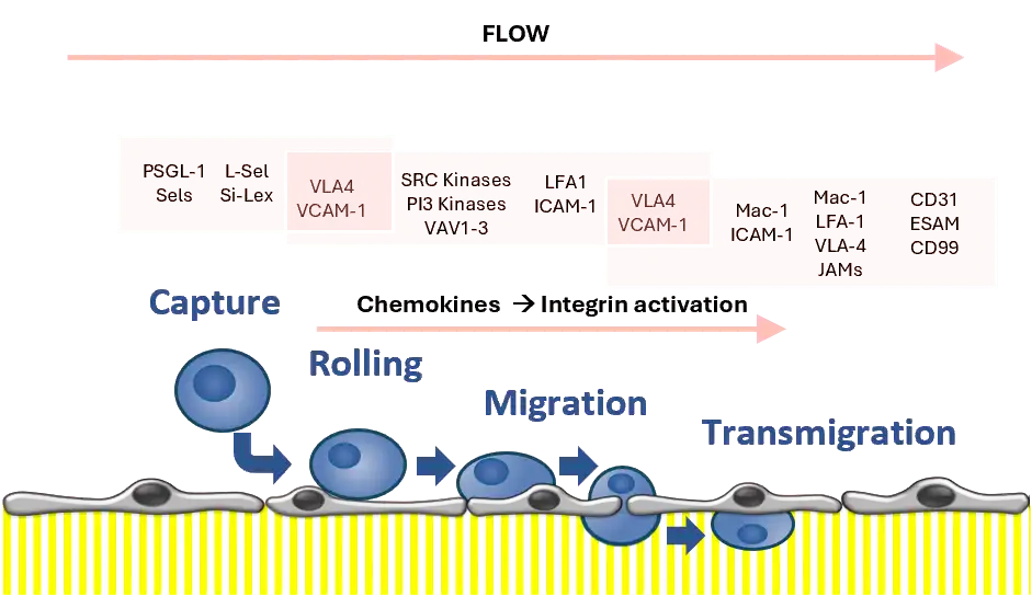
Leukocytes captured and rolling on endothelial cells
➔
Leukocytes transmigrated through endothelial cells
Standard bio-imaging time course of leukocytes, where cells would typically be counted manually.
MesenCount in action: red squares mark the migrating cells, while purple squares mark the transmigrated cells through vascular endothelium.
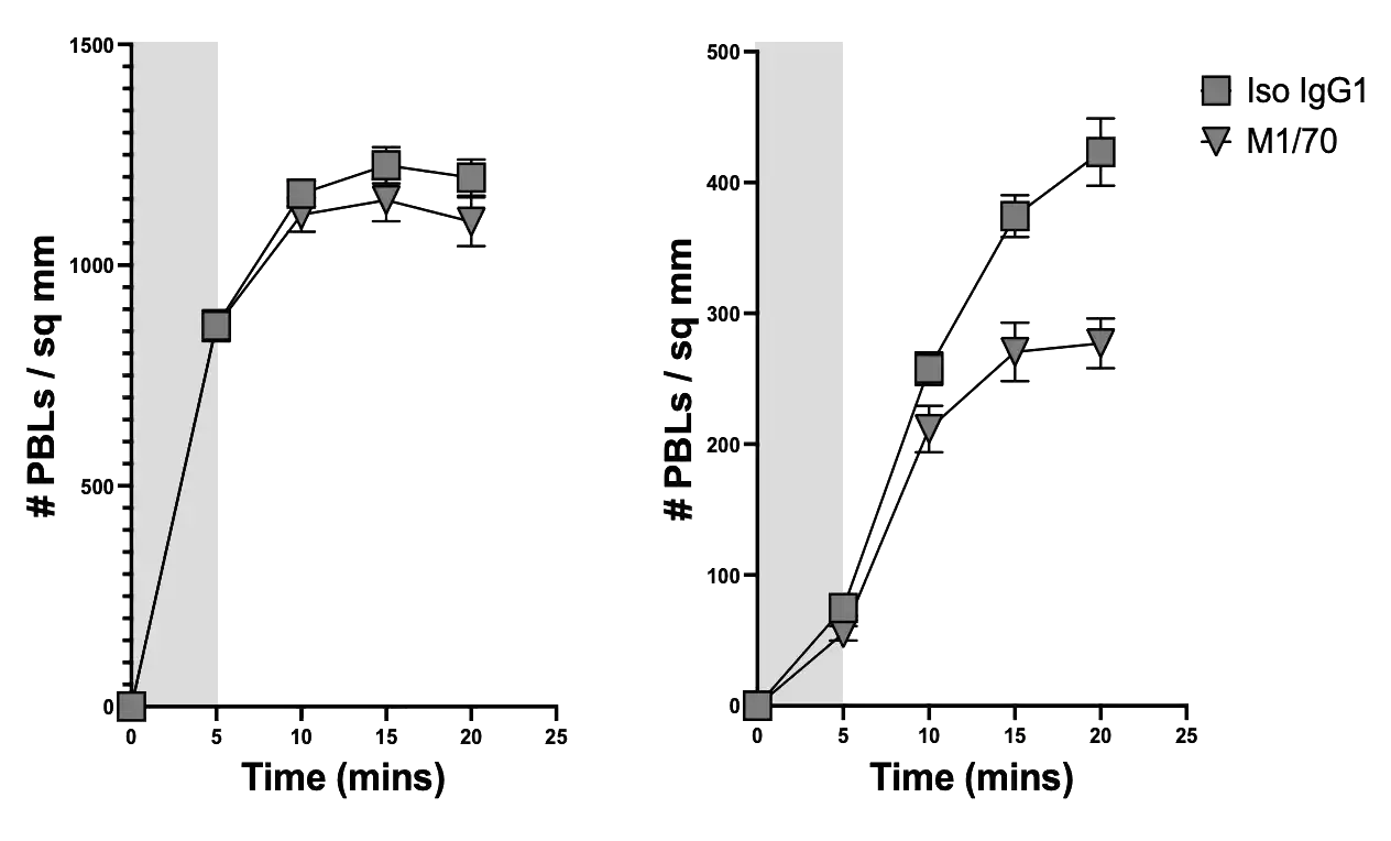
Time-course analysis
1) Capture
2) Transmigrated
Area under curve (AUC) analysis
1) Capture
2) Transmigrated
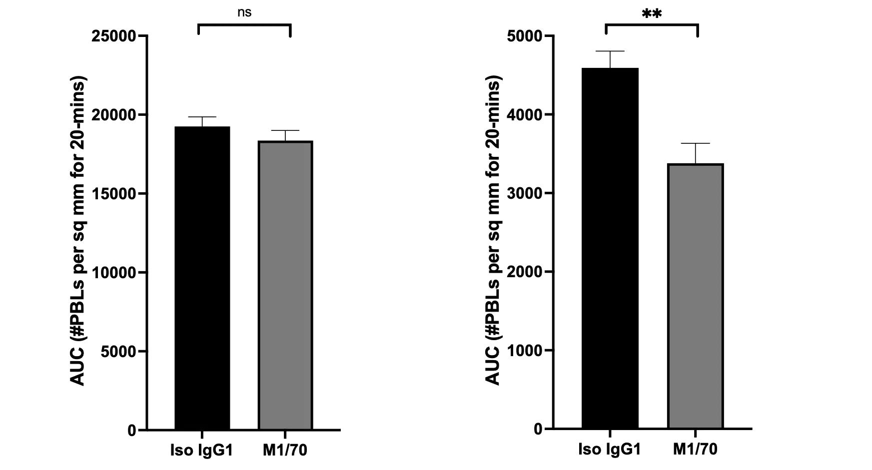
Fixed triplicate fields at 5-min intervals.
Mean + standard error
Statistics: Student T-Test
ns: not significant; **: P 0.01
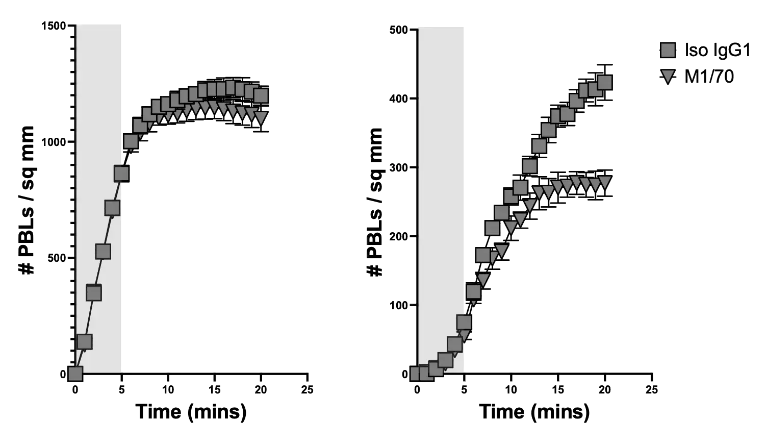
Time-course analysis
1) Capture
2) Transmigrated
Area under curve (AUC) analysis
1) Capture
2) Transmigrated
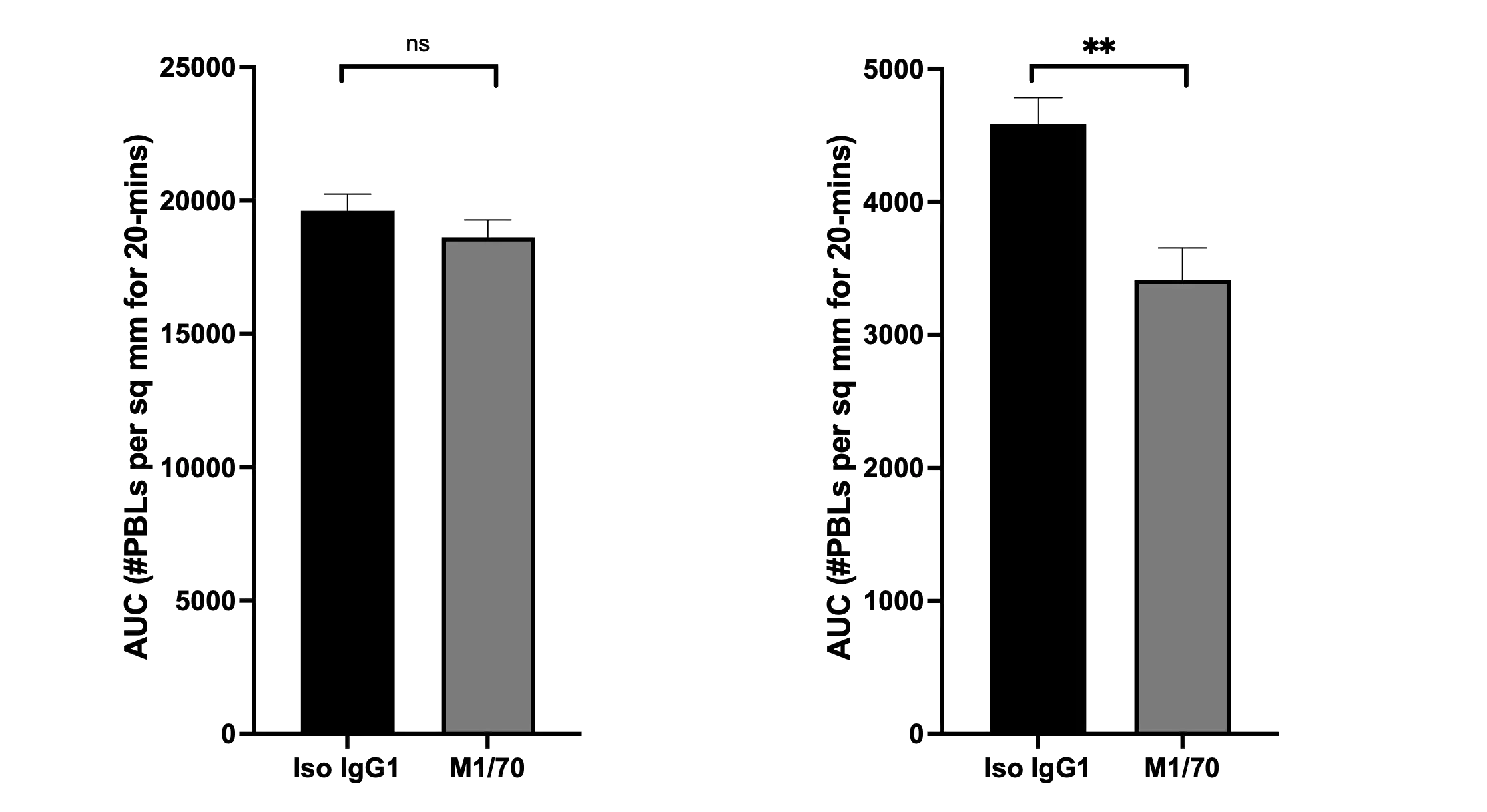
Fixed quadruplicate fields at 1-min intervals.
Mean + standard error
Statistics: Student T-Test
ns: not significant; **: P 0.01
3) Cell speed
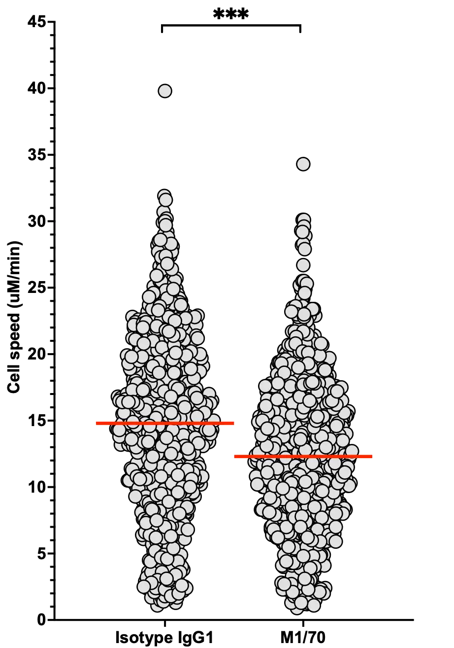
Profile of human peripheral blood leukocytes (PBLs) trafficking in human umbilical vein endothelial cells (HUVECs) under flow.
PBLs were flowed over activated HUVECs for 5 minutes (area marked in grey). Captured PBLs were individually tracked, and their positions marked at set intervals. PBLs captured from free flow became rapidly activated by rolling on the luminal surface, changing phase from white to grey (Capture) before transmigrating into the ablumen (Transmigration).
MesenFlow Technologies SàrlChemin des Aulx 14
1228 Plan-les-Ouates
Geneva, Switzerland
+41 22 32 16 961 (office)
+41 79 36 66 291 (mobile)