Our vascularized organoid model recreates complex tissue microenvironments to study leukocyte trafficking and endothelial interactions. By integrating proprietary tumor or organoid context, it enables deeper insights into cell behavior and improves the predictive power of preclinical studies.

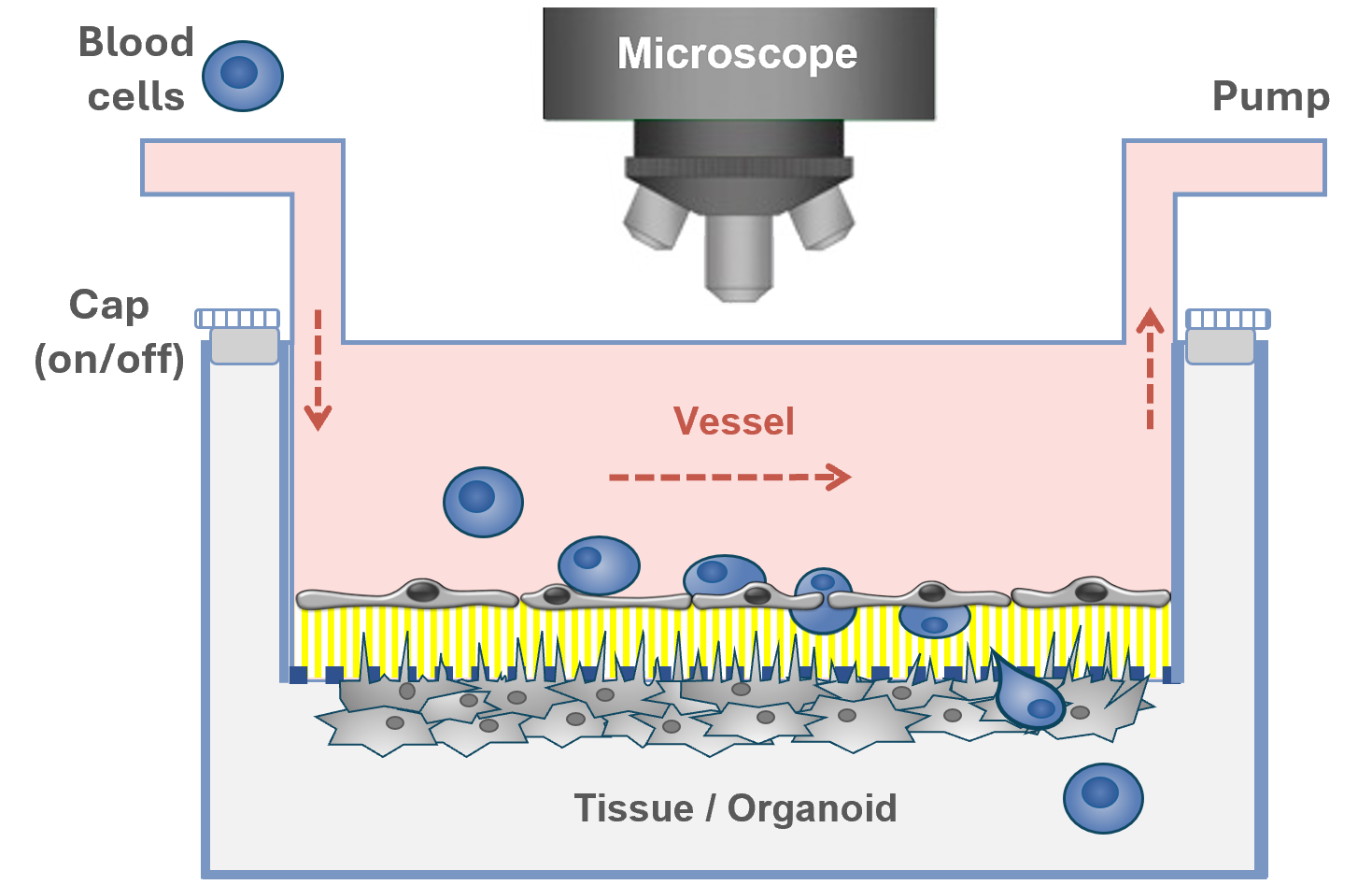
Tissue and vascular modeling
› Micro-physiological system (MPS) using dual-chamber design
› Vascular flow: capture, rolling, migration and transmigration

Organ-specific drug testing
› Replicating complex physiological interactions
› Blood-brain-barrier (BBB) model (drug penetration)
› Other organoid models on request

Mechanism of action (MoA)
› Analysis of endothelial activation pathways
› Measurement of adhesion molecules and cytokines
› Identification of pro- or anti- inflammatory effects

Bio-imaging
› High-resolution imaging by phase contrast microscopy
› Immunohistochemistry: visualisation of cellular morphology, drug-specific effects
› Adjustable time-course measurements

Cell tracking
› MesenCount and MesenTrack AI-based softwares
› Label-free or immunofluorescence imaging
under flow

Flow Cytometry
› Recovery and phenotyping of recruited leukocyte subsets in whole blood models
› Identification of endothelial cell activation markers
› Measurement of cytokines production
Example 1
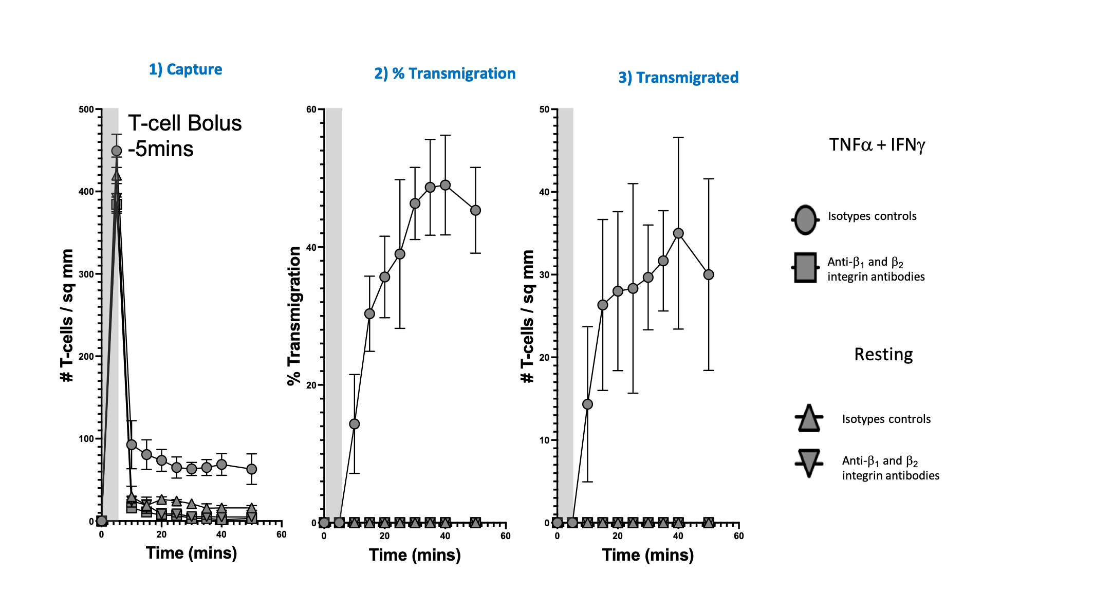
Trafficking profiles of T-cells on resting human induced pluripotent stem cell-derived brain microvascular endothelial cells (iBMECs) or stimulated with TNFα + IFNγ. The number of T-cells captured (step-1), the percentage transmigration (step-2) and the number of transmigrated T-cell (step-3) were monitored over 50 minutes, with individual cells quantified using our AI-based software MesenCount. Treatment with anti-β1 and anti-β2 integrin antibodies (square and inverted triangle) markedly reduces T-cell capture and transmigration compared to the isotype control (circle and upright triangle).
Example 2
Dual-chamber flow system
3µM pore PET filter
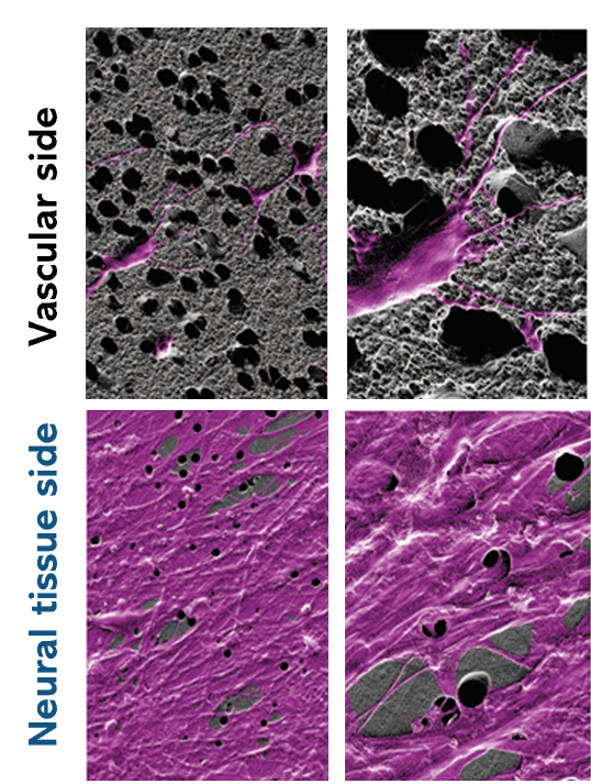
Cell-cell contact between both chambers
Electron micrograph of PET filter
Neural tissue marked in purple
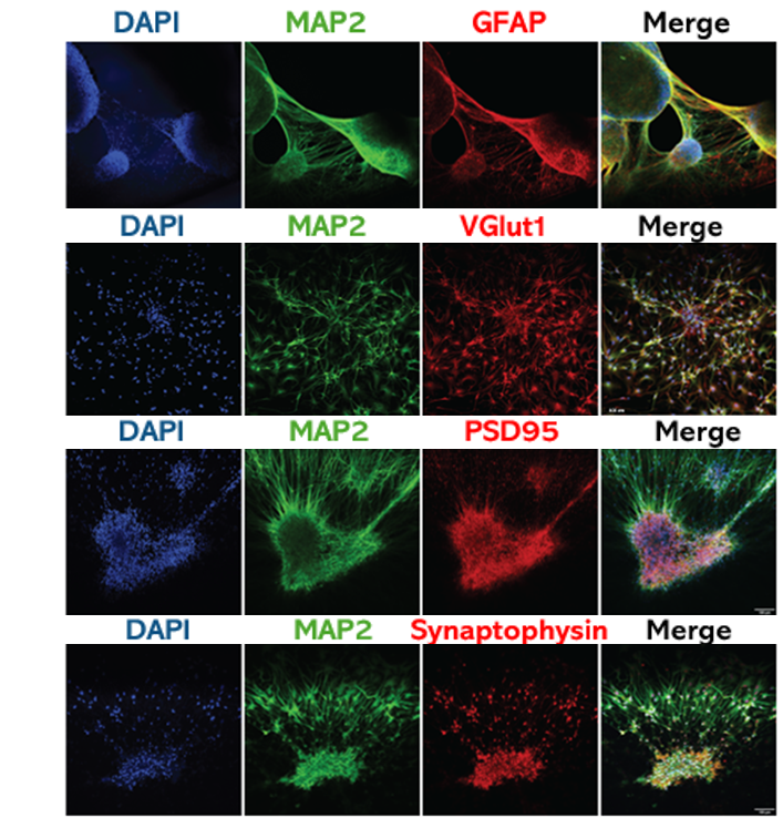
2D neural tissue –
Validation makers at 7 days of maturation
Dual-chamber flow system employing 2D neural tissue. Scanning electron microscopy confirmed thetissue protrudes through the membrane, indicating physical contact between the endothelium andneural tissue compartments. Cultured neural tissue express neuronal, glial and synaptic markers.
Images data generated by Hepia


MesenFlow Technologies SàrlChemin des Aulx 14
1228 Plan-les-Ouates
Geneva, Switzerland
+41 22 32 16 961 (office)
+41 79 36 66 291 (mobile)