Our inflammation model recreates acute or chronic inflammatory states in human endothelial cells. This platform enables evaluation of compound efficacy and a reliable way to study inflammatory responses in a vascular micro-environment.
Acute persistent inflammation
› Non-alcoholic Steatohepatitis (NASH)
› Western Diet Obesity
› Atherosclerosis
› Diabetes-II
Chronic non-resolving inflammation
› Multiple Sclerosis
› Rheumatoid Arthritis
› Inflammatory Bowel Disease
Vascular pathologies
› Sickle Cell Disease
› Vascular Occlusion
› Ischemia

Anti-inflammatory drug development
› Therapeutic and prophylactic protocols available
› Multi-throughput screening
› Dose response analysis and IC50 determination

Mechanism of Action (MoA) studies
› Analysis of endothelial activation pathways
› Measurement of adhesion molecules and cytokines
› Identification of pro- or anti-inflammatory effects

Vascular modeling
› Regular vascular flow: leukocytes trafficking measurements (capture, rolling, migration and transmigration)
› Disruptive flow: monocytes or neutrophils platelet aggregates
› Rolling leukocyte flow: endothelial cells replaced by platelets

Chronic and acute disease modeling
› Acute-persistent inflammation:
• Neutrophil extra cellular traps (NETs)
• Oxidized low-density lipoprotein (OxLDL)
• Tumor necrosis factor superfamily (TNFSF14)
› Chronicnon-resolving inflammation:
• Tumor necrosis factor alpha (TNFα)
• Interfer on gamma (IFNɣ)
• Lipopolysaccharides (LPS)

Safety and toxicity assessment
› Endothelial cell activation and dysfunction
› Endothelial junctions integrity

Bio-imaging
› High-resolution imaging
by phase contrastmicroscopy
› Immunohistochemistry: visualisation of cellular morphology, drug-specific effect
› Adjustable time-course measurements

Cell tracking
› MesenCount and MesenTrack AI-based softwares
› Label-free or immunofluorescence imaging
under flow

Flow Cytometry
› Recovery and phenotyping of recruited leukocyte subsets in whole blood models
› Identification of endothelial cell activation markers
› Measurement of cytokines production
Example 1
Trafficking of human monocytes on activated HUVECs under flow
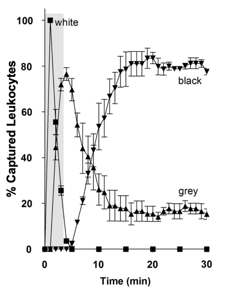
Trafficking of human monocytes on activated HUVECs under flow. Monocytes flowed over HUVECs for 5-mins (area marked in grey). They were tracked, andtheir positions marked at 1-min intervals. Monocytes captured from free-flow became rapidly activated by rolling on the luminal surface, changing phase from white(squares) to grey (triangles). Monocytes that underwent transmigration into the ablumen changed to phase-black (inverted triangles).
Example 2
Blockade in Monocyte transmigration identified with Compound-C
Success with a typical screening strategy
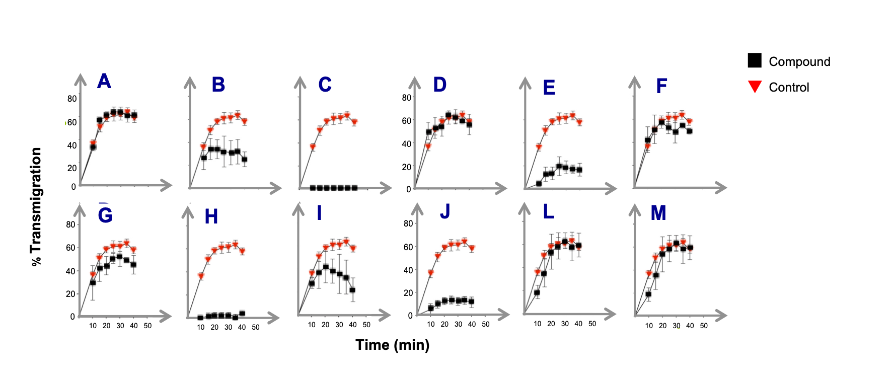
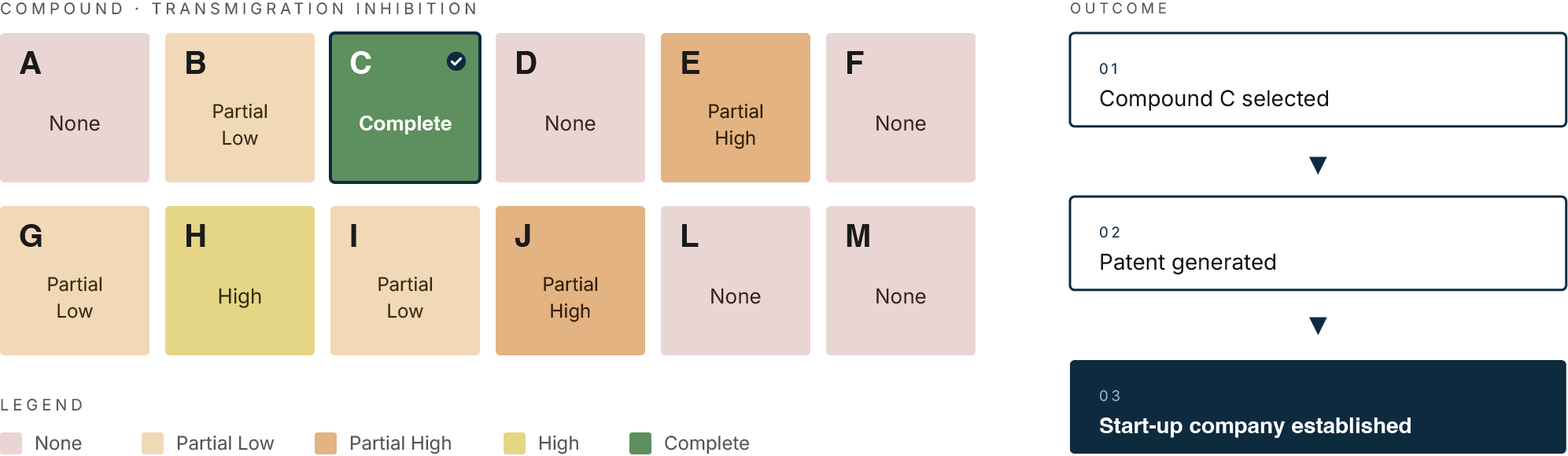
Monocyte transmigration on the surface of activated HUVECs under flow. Primary human monocytes co-cultured on TNFα-activated HUVECs. Test compounds at 30 µM or control were added to cells for 45 min and the transmigration blockade recorded.
MesenFlow Technologies SàrlChemin des Aulx 14
1228 Plan-les-Ouates
Geneva, Switzerland
+41 22 32 16 961 (office)
+41 79 36 66 291 (mobile)